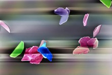
We are pleased to announce the winners of the 2015 School of Science Research Photo Contest. This year’s exceptional entries demonstrated the broad scope of research being conducted within the School as well as the creative skills required to capture the nature of the research. Thank you to all who participated in the contest and congratulations to our winners!
Grand Prize Winner
"Mother Nature's Spring Origami"
by Yunbo Yang
Description:The nanoscale butterfly (top), flower and bird (bottom left), petals and leaves are made from layered material. Material in the photo: SnS2 grown by co-evaporation of Sn and sulfur.
Category Winners:
Data Analytics & Predictive Modeling:
“Active Gene Connections Compared Across 6 Brain Diseases” by Matthew Poegel
Description: The results of data analysis on the activity of genes associated with six brain diseases--Alzheimer's, Autism, Holoprecencephaly, Lissencephaly, Microcephaly, and Tauopathy--have revealed six clusters representing different stages of brain development. The connections between genes illustrate links between them while the outer heat map tracks the activity of the genes over nine intervals during a period of 77 days.
Water, Energy, Resilience, and Sustainability:
“Gray Treefrog” by Brian Mattes
Description: My lab has done a lot of work with amphibians and phenotypic plasticity. The Gray Treefrog (Hyla versicolor) has been studied and found to exhibit amazing responses to natural and anthropogenic factors. Predators can induce a color change in the tails of tadpoles, and small exposures to pesticides can effect tolerance to that pesticide later in life.
Computational Science, Security, and Simulation:
“Water Desalination” by Adrien Nicolaï
Description: Water desalination using nanostructured porous materials. These simulations at CCI reveal the potential of graphene oxide frameworks, pictured in black, to remove contaminants such as salt ions, seen in blue and green, from water.
Biomedical Science and Applications:
“Human Neural Development” by Greg Nierode
Description: This contains an immunofluorescence image depicting human neural development. It shows human neural stem cells that have differentiated into neurons (green) and astrocytes (orange). The nuclei of the cells are colored blue.
Materials at the Nanoscale:
“Poly-l-lactide Composite Cross-section” by Kristen Lee
Description: Scanning electron micrograph of a poly-l-lactide extruded microfiber with an electrospun nanofiber coating.
All winning photos may be viewed at the School of Science Photobucket page.
